Spatial Profiling developing deep datasets from single tissue sections
Spatial profiling allows researchers to view and quantify molecular markers and cell types in the context of architecturally intact tissue sections — capturing information about marker co-expression, tissue composition
( heterogeneity ), and tumor microenvironment organization.
( heterogeneity ), and tumor microenvironment organization.
Instruments
GeoMx Digital Spatial Profiler (DSP)
Technologic innovations and new imaging platforms are shaping the field of spatial biology.
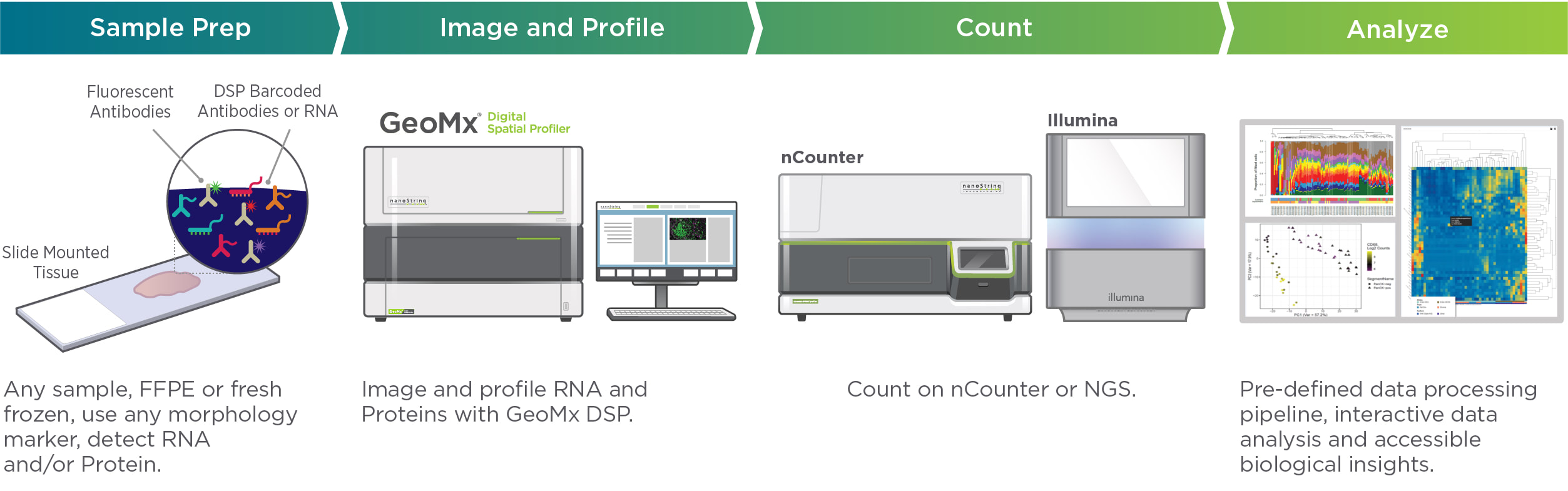
The Dana-Farber Pathology Core lab currently uses NanoString GeoMx Digital Spatial Profiler (DSP) digital optical barcoding to perform highly multiplexed, spatially resolved profiling experiments.
Up to 4 fluorescent antibodies guide selection of regions of interest (ROI).
Selected regions are then profiled for up to thousands of RNAs or scores of protein biomarkers using panels of barcoded in situ probes or antibodies from Nanostring. These panels can be customized to probe for RNA sequences of interest or to include antibodies selected by your team. Barcodes attached to the RNA probes or antibodies by photocleavable linkers are cleaved and 'lifted' from ROIs and then sequenced for counting.
Investigators will work closely with Core staff and Nanostring representatives to design experiments. Research teams should include collaborating pathologists as well as data analysis teams.
Overview of the Nanostring GeoMx Workflow
For more information about GeoMx technology, visit the Nanostring website.
Be sure to look at predesigned Nanostring oncology panels which interrogate critical cancer cell signaling pathways and markers of inflammatory signatures in tumors.
Be sure to look at predesigned Nanostring oncology panels which interrogate critical cancer cell signaling pathways and markers of inflammatory signatures in tumors.